Electron Microscopy at the University of
Tampa
In
the summer of 2011, the University of Tampa purchased and installed a JEOL
6010LA Analytical Scanning Electron Microscope to support its program in electron
microscopy. This instrument will serve as the focus for formal courses
(Essentials of Electron Microscopy and Forensic Science) as well as a workhorse
for faculty and student research. Below are some of the first images obtained
with this new instrument. Updated 5/5/2016
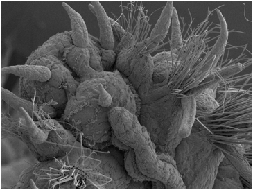
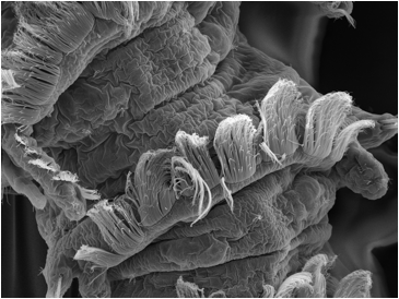
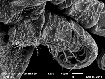
Fireworm head
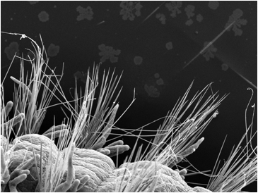
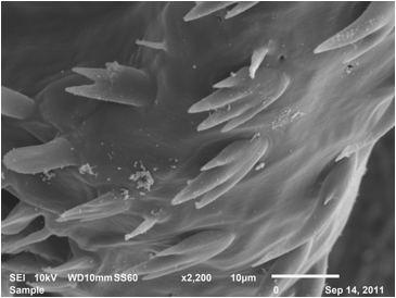
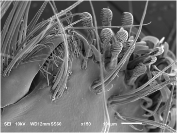
Fireworm setae
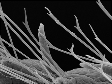
Close-up of Fireworm setae
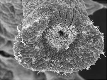
Terminal segment of the local
Polychaete, Polydora
Dorsal cilia of Polydora
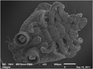
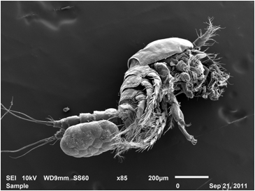
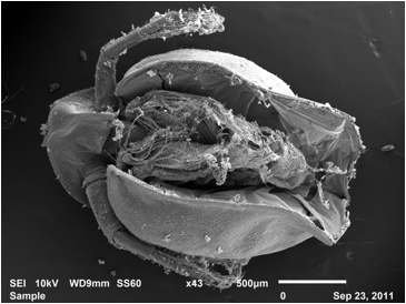
Ventral surface of the fish
louse, Argulus
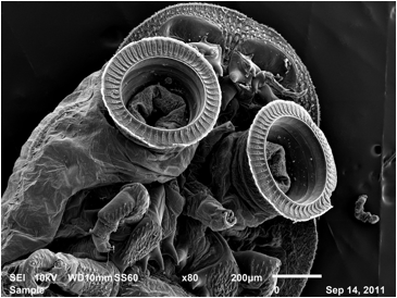
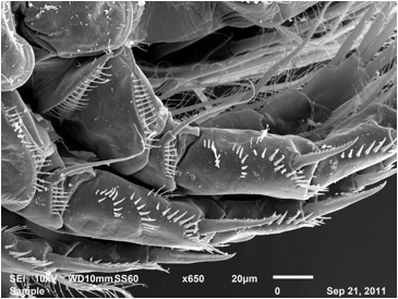
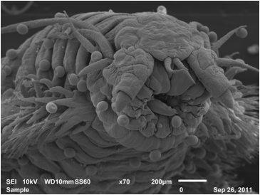
Ventral suckers of Argulus
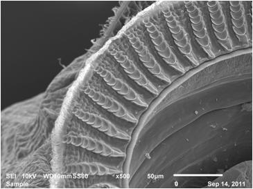
Detail of the Argulus sucker
Thoracic leg of Argulus
Microspines on Argulus
Female Copepod with egg
case
Copepod leg spines
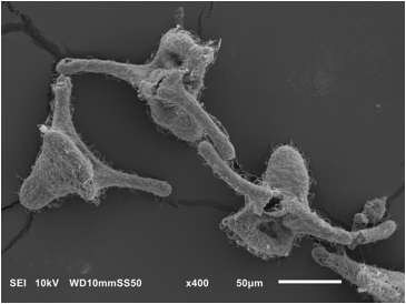
Clausidium female with attached male
Clausidium female with attached male
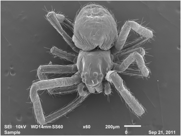
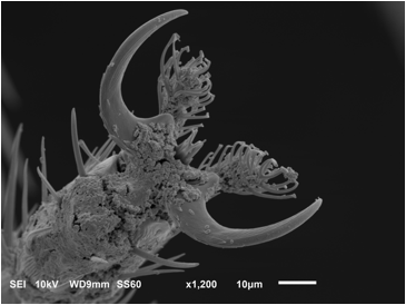
Brown Widow Spider Juvenile
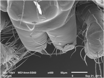
Brown Widow Spider fangs
Sanddollar spine
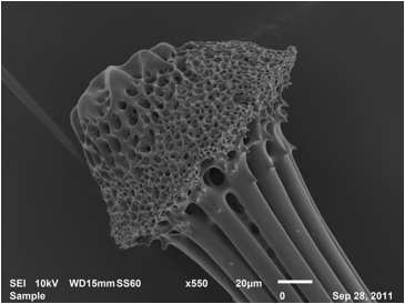
Base of Sanddollar
spine
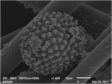
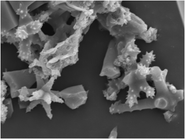
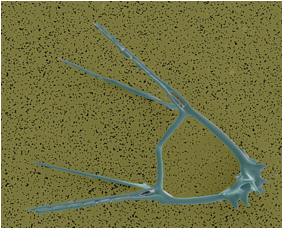
Sponge Spicules
Sponge Spicules
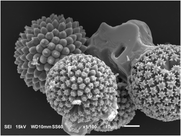
Sponge Spicules
Sponge Spicules
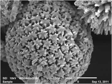
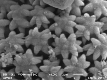
Small Sponge Spicules (5500x)
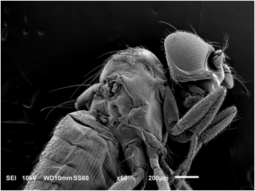
Wingless Mutant of Drosophila
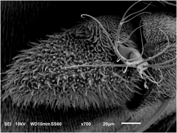
Drosophila
antenna
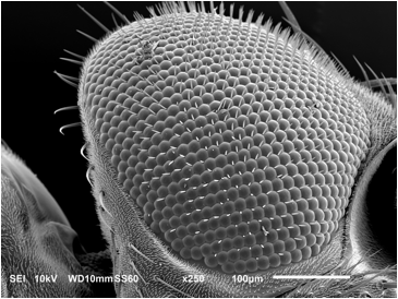
Drosophila
eye
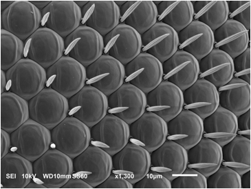
Drosophila
eye units (ommatidia)
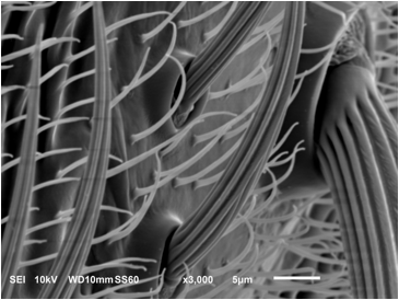
Drosophila
setae
Drosophila
toes (tarsal spines)
Daphnia, a
Freshwater Crustacean
Nereid polychaete
head
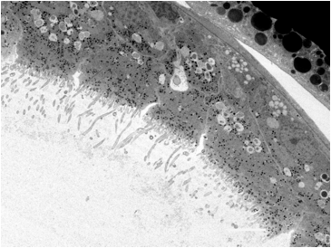
The STEM (Scanning
Transmission Electron Microscope) adaptor allows us to view thin sections on
the SEM
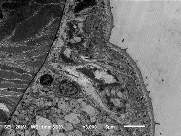
Section of epidermal glands
in the polychaete, Streblospio
Section of the gut lining in Streblospio
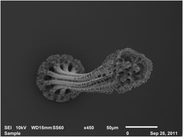
Pluteus larvae of Sea Urchin
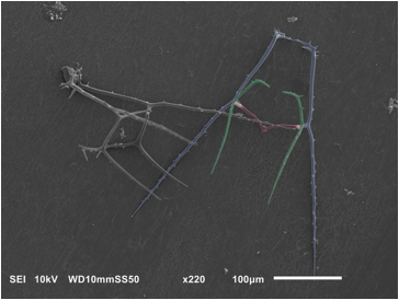
Lytechinus
larval skeletons
Arbacia
larval skeletons
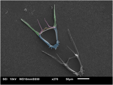
Arbacia 7 day larval skeleton

Lytechinus 11 day larval skeleton
Breaking News! The ubiquitous
pasta scoop
was discovered by Stomatopods!